10,000 pg/mL: Add 1 mL of pattern diluent buffer into one tube of normal (10 ng per tube) and blend completely. Note: Store this resolution at 4°C for as much as 12 hours (or -20°C for 48 hours) and keep away from freeze-thaw cycles.
5,000 pg/mL: Mix 0.Three mL of 10,000 pg/mL with 0.Three mL of pattern diluent buffer and blend completely.
2,500 pg/mL: Mix 0.Three mL of 5,000 pg/mL with 0.Three mL of pattern diluent buffer and blend completely.
Perform comparable dilutions till the usual options with these concentrations (pg/mL) are made:
1,250, 625, 312, 156 and 78.
Add 100 µL of every of the diluted commonplace options to the suitable empty wells. Repeat in duplicate or triplicate for accuracy. Note: The commonplace options are finest used inside 2 hours.
Biotinylated Antibody
Calculate the overall quantity wanted for the assay by multiplying 0.1 mL/effectively and the variety of wells required. Add 2-Three additional wells to the calculated variety of wells to account for doable pipetting errors.
Generate the required quantity of diluted antibody by performing a 1:100 dilution (For every 1 µL concentrated antibody, add 99 µL antibody dilution buffer) and mixing completely.
Avidin-Biotin-Peroxidase Complex (ABC)
Calculate the overall quantity wanted for the assay by multiplying 0.1 mL/effectively and the variety of wells required. Add 2-Three additional wells to the calculated variety of wells to account for doable pipetting errors.
Generate the required quantity of diluted ABC resolution by performing a 1:100 dilution (For every 1 µL concentrated ABC resolution, add 99 µL ABC dilution buffer) and mixing completely. Note: The diluted ABC resolution shouldn’t be ready greater than 1 hour previous to the experiment.
Sandwich ELISA ProtocolAll the ELISA kits from Boster use the sandwich format and biotin-streptavidin chemistry. Our ELISA assays require the dilutions of normal options, biotinylated antibody (detection antibody) and avidin-biotin-peroxidase complicated.
Capture Antibody Coating(These steps aren’t required if the pre-adsorbed Picokine ELISA kits from Boster are used)
Dilute the seize antibody to a final focus of 1-10 μg/mL in bicarbonate/carbonate antigen-coating buffer (100 mM NaHCO3 in deionized water; pH adjusted to 9.6).
Pipette 100 μL of diluted antibody to every effectively of a microtiter plate.
Cover the plate with adhesive plastic and incubate at 4°C in a single day (or 37°C for 30 min).
Remove the coating resolution and wash the plate 3X with 200 μL PBS (Phosphate Buffered Saline) buffer (10 mM Na2HPO4 and 1.eight mM NaH2PO4 in deionized water with 0.2% Tween 20; pH Adjusted to 7.4) with for five minutes every time. The coating/washing options may be eliminated by flicking the plate over a sink. The remaining drops may be eliminated by patting the plate on a paper towel or by aspiration. Do not enable the wells to dry out at any time.
Blocking(These steps aren’t required if the pre-adsorbed Picokine ELISA kits from Boster are used)
Pipette 200 μL blocking buffer (5% w/v non-fat dry milk in PBS buffer) per effectively to dam residual protein-binding websites. Alternatively, BSA or BlockACE can be utilized to switch non-fat dry milk.
Cover the plate with adhesive plastic and incubate for 1-2 hour(s) at 37°C (or at 4°C in a single day).
Remove the blocking resolution and wash the plate 2X with 200 μL PBS for five minutes every time. Flick the plate and pat the plate as described within the coating step.
Reagent Preparation
Prepare for the diluted commonplace options, biotinylated antibody and ABC options as proven within the above Reagent Preparation part. Note: The diluted ABC resolution shouldn’t be ready greater than 1 hour previous to the experiment.
Sample (Antigen) Incubation
Serially dilute the pattern with blocking buffer instantly earlier than use. The optimum dilution ought to be decided by a titration assay in accordance with the antibody producer.
Pipette 100 μL of every of the diluted pattern options and management to every empty effectively. Repeat in duplicate or triplicate for accuracy. The destructive management ought to be species- and isotype-matched in addition to non-specific immunoglobulin diluted in PBS buffer.
Cover the plate with adhesive plastic and incubate for two hours at room temperature.
Remove the content material within the wells and wash them 3X with 200 μL PBS buffer for five minutes every time. Flick the plate and pat the plate as described within the coating step.
Biotinylated Antibody Incubation
Pipette 100 μL of diluted antibody to the wells with management, commonplace options and diluted samples.
Cover the plate with adhesive plastic and incubate for 1 hour at 37°C (or 2 hours at room temperature). These incubation instances ought to be adequate to obtain a robust sign. However, if a weak sign is noticed, carry out incubation in a single day at 4°C for a stronger sign.
Remove the content material within the wells and wash them 3X with 200 μL PBS for five min every time. Flick the plate and pat the plate as described within the coating step.
ABC Incubation
Pipette 100 μL of diluted ABC resolution to the wells with management, commonplace options and diluted samples.
Cover the plate with adhesive plastic and incubate for 0.5 hour at 37°C.
Remove the content material within the wells and wash them 3X with 200 μL PBS buffer for five min every time. Flick the plate and pat the plate as described within the coating step.
Substrate PreparationPrepare the substrate resolution instantly earlier than use or convey the pre-made substrate to room temperature. The two broadly used enzymes for sign detection are horse radish peroxidase (HRP) and alkaline phosphatase (AP), and their corresponding substrates, stopping options, detection absorbance wavelengths and colour developed are as follows:EnzymeSubstrate*Stop SolutionAbsorbance (nm)Color DevelopedHRPTMB2M H2SO4450YellowAPpNPP0.75M NaOH405Yellow* TMB: 3,3’,5,5’-tetramethylbenzidine; pNPP: p-nitrophenyl-phosphate Note:
The TMB substrate should be saved at 37°C for 30 min earlier than use.
Hydrogen peroxide may also act as a substrate for HRP.
Sodium azide is an inhibitor of HRP. Do not embrace the azide in buffers or wash options if HRP-labeled conjugate is used for detection.
Signal Detection
Pipette 90 μL of substrate resolution to the wells with the management, commonplace options and diluted samples.
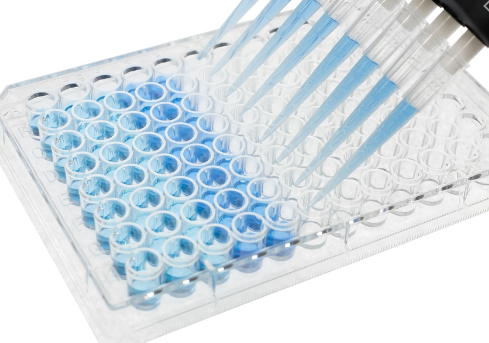
Incubate the plate at 37°C at the hours of darkness. If TMB is used, shades of blue can be noticed within the wells with essentially the most concentrated options. Other wells could present no apparent colour.
Color ought to be developed in constructive wells after 15 min. After adequate colour growth, pipette 100 μL of cease resolution to the suitable wells (if obligatory).
Read the absorbance (OD: Optical Density) of every effectively with a plate reader.
Data Analysis
Prepare a normal curve utilizing the information produced from the diluted commonplace options. Use absorbance on the Y-axis (linear) and focus on the X-axis (log scale).